If you're looking for a dentist near me in Walnut Creek, CA, there's a good chance you're also wondering what a new patient visit will feel like. For many people, the word "X-rays" brings up old memories of awkward film tabs, waiting around, and not quite knowing what the dentist is seeing.
That experience has changed.
At a modern dental office, digital dental imaging helps make visits faster, clearer, and easier to understand. Instead of taking an image and disappearing to develop film, your dental team can often bring the image up on a screen right away and walk you through what it means. That matters whether you're coming in for cleaning and exams, dealing with tooth pain, exploring dental implants near me, or looking for a cosmetic dentist near me.
For patients in Walnut Creek and nearby East Bay communities, this technology isn't just about newer equipment. It's about feeling more informed, more comfortable, and more confident in your care.
A Clearer Picture of Your Health with Your Dentist in Walnut Creek
When you come in for a routine exam, a problem-focused visit, or an emergency dentist appointment, your dentist needs a view that goes beyond what the eye can see. Teeth can look healthy on the surface while decay starts between them. A sore tooth can feel obvious to you, but the reason behind that pain may sit under the gumline or around the root.
That's where digital dental imaging helps.
Why modern imaging matters in daily dental care
Digital images give your dentist a practical, everyday way to check for issues early and plan treatment with more confidence. In a family practice setting, that can support decisions about preventive care, fillings, crowns, root canals, tooth extraction, and restorative dentistry.
This isn't a niche technology anymore. The global dental imaging market was valued at USD 2.28 billion in 2025 and is projected to reach USD 4.71 billion by 2034, with North America identified as the largest regional market in that forecast, according to Fortune Business Insights on the dental imaging market. For patients, the takeaway is simple. Digital imaging has become a mainstream part of modern dental diagnosis and treatment planning.
What this means for you: when a dental office uses digital imaging as part of everyday care, you're seeing a tool that's now central to how many dentists evaluate problems and explain treatment.
What patients usually notice first
Most patients don't care about the technical terms at first. They care about whether the appointment feels smooth and whether the explanation makes sense.
Digital imaging helps with both:
- Faster viewing: images appear quickly, so there isn't the same delay associated with older film systems.
- Clearer discussion: your dentist can point to a specific tooth, root, or area of bone on a monitor while explaining what's going on.
- Better planning: if you're considering cosmetic dentistry, restorative dentistry, or implants, imaging helps turn a vague conversation into a specific plan.
If you've been putting off a visit because you're worried about discomfort, time, or uncertainty, digital imaging often makes the process feel much more approachable. That's a big reason many patients who are searching for a dentist in Walnut Creek, CA ask about technology before they book.
What Is Digital Dental Imaging
Think of the change from film X-rays to digital imaging the same way you might think about old film cameras versus today's digital cameras. With film, you had to capture the picture, process it, and wait. With digital, the image appears quickly and can be enlarged, adjusted, and reviewed right away.
That's the basic idea behind digital dental imaging.
How the image becomes visible
Inside the equipment, a receptor captures the X-ray signal and converts it into digital information. Dental imaging systems commonly operate at 8-, 10-, 12-, or 16-bit depth, and that added bit depth improves grayscale precision and gives the dentist more flexibility when adjusting contrast, zoom, and edge detail, as described in this peer-reviewed review of digital image processing in dental radiology.
If that sounds technical, here's the plain-English version: the software can help your dentist see fine differences in shades of gray. That can make small areas easier to evaluate, including subtle changes near a tooth root or areas that aren't obvious during a visual exam alone.
A digital image isn't "guessing." It's converting what the sensor captures into a format your dentist can enlarge, review, and explain chairside.
Common types of digital images
Different kinds of dental visits call for different views.
Intraoral images
These are the close-up images taken with a small sensor inside your mouth. They help your dentist examine individual teeth, spaces between teeth, fillings, and areas around the roots.
A patient coming in for a new patient exam, sensitivity, or possible cavity often starts here.
Panoramic images
A panoramic image gives a wider view of your full mouth in one scan. It can help your dentist evaluate tooth position, jaw structure, and broader patterns that don't show up on a close-up image alone.
This is often useful in thorough exams, wisdom tooth evaluation, and treatment planning.
CBCT scans
Cone Beam Computed Tomography, often called CBCT, creates a 3D view. That extra detail can be especially helpful when your dentist needs to study bone, root position, or anatomical structures before more involved treatment.
Patients usually hear about CBCT when discussing implants, complex extractions, or other advanced planning needs.
Why this matters to patients
You don't need to memorize the types. What matters is that your dentist can choose the right view for the right problem. The result is a more precise conversation about what you're feeling, what the image shows, and what the next step should be.
The Benefits of Digital X-Rays for Your Family
When patients ask why a dental office uses digital X-rays instead of film, the answer isn't "because it's newer." The answer is that it improves the patient experience in ways families can actually feel.
Lower radiation is the first reason many patients ask
One of the biggest advantages is reduced radiation exposure. Peer-reviewed research notes that digital radiography can reduce dose by up to 80% versus conventional film, and some modern systems may reduce exposure by as much as 90% compared with older film X-rays, according to this review of digital radiography in dentistry.
For parents, safety-conscious adults, and patients who need periodic imaging, that's often the most reassuring part of the conversation.
Practical rule: if imaging is needed to diagnose a problem or plan treatment, patients usually want two things at once. They want the image to be useful, and they want the process to be as safe as possible.
Other benefits patients notice during the visit
Digital X-rays also make appointments feel more efficient and collaborative.
- Quicker appointments: images are available without film development, so there is less waiting.
- Better patient education: your dentist can enlarge an image and show you the exact area being discussed.
- Simpler record handling: digital files are easier to store and retrieve, which supports continuity of care.
- Cleaner workflow: digital systems avoid the chemical film processing associated with older methods.
Digital Imaging vs. Traditional Film X-Rays
| Feature | Digital Imaging (Our Practice) | Traditional Film X-Rays |
|---|---|---|
| Image availability | Appears quickly on a computer screen | Requires film processing |
| Radiation exposure | Lower exposure in many systems, including up to 80% less than conventional film in peer-reviewed review, with some modern systems reporting as much as 90% less than older film X-rays | Higher exposure than digital systems described above |
| Image review | Can be enlarged and adjusted for contrast and detail | More limited post-processing |
| Storage | Electronic storage and retrieval | Physical film handling and storage |
| Patient conversation | Easy to review together chairside | Less interactive in many settings |
Why this helps in family and preventive care
For routine dental care, preventive visits, and new patient exams, digital imaging supports earlier discussions about what your dentist sees and what needs attention now versus later. That can be especially helpful if you want to stay ahead of small issues instead of waiting until pain pushes you into an urgent visit.
How We Use Imaging for Dental Implants and Cosmetic Dentistry
A good image becomes even more valuable when treatment is more involved than a routine checkup. That's where patients often see the practical side of digital imaging most clearly.
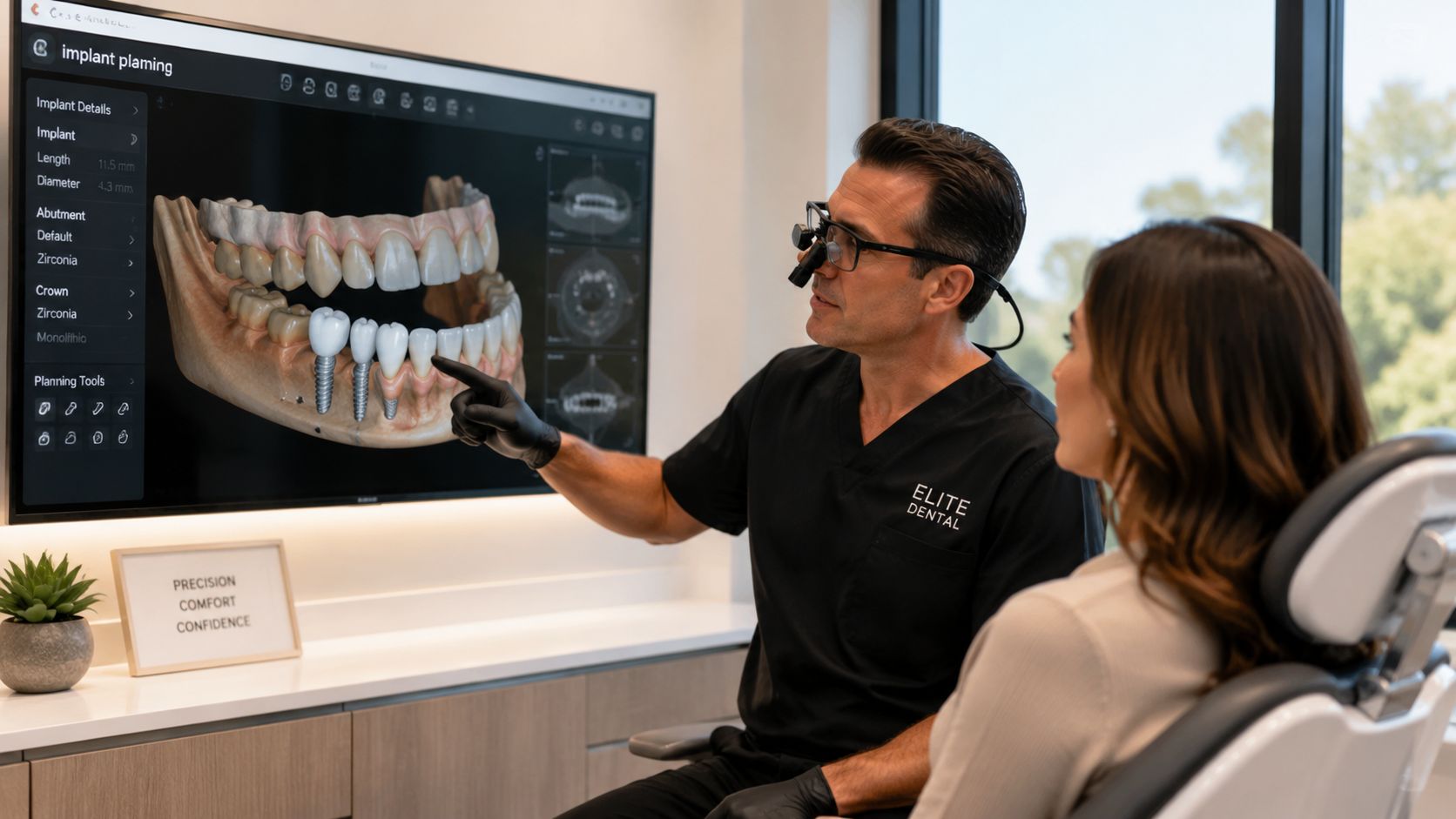
For dental implants
Say a patient is missing a tooth and searches for dental implants near me. They don't just want a replacement. They want to know whether the implant will fit well, function well, and look natural.
That planning starts with the right image. A 3D scan can help your dentist evaluate available bone, see important structures, and map out implant placement before treatment begins. If you're exploring dental implants in Walnut Creek, CA, digital imaging helps turn the process into a careful, visual plan rather than a rough estimate.
A patient often feels calmer once they can see the area on a screen and understand why a certain approach makes sense.
For cosmetic dentistry
Cosmetic work is personal. If you're thinking about veneers, teeth whitening, or other smile improvements, you want your dentist to pay attention to detail.
Digital images help your dentist study tooth shape, spacing, symmetry, and how existing dental work appears alongside natural teeth. That matters when planning cosmetic improvements that should look balanced, not artificial.
Patients searching for a cosmetic dentist near me usually want two things. They want a smile that looks better, and they want confidence that underlying dental health has been checked first. Imaging supports both goals.
The best cosmetic result starts with a clear diagnosis. A beautiful smile still has to function comfortably and predictably.
For root canals, restorative dentistry, and difficult symptoms
Digital imaging also helps when the problem isn't appearance at all.
A patient may come in with sharp pain when biting, swelling, or a tooth that feels "off" but doesn't show an obvious crack from the outside. In those cases, imaging helps your dentist evaluate the roots, surrounding bone, and prior dental work.
That can guide decisions about:
- Root canal treatment: when a deeper infection or inflammation may be involved
- Crowns and bridges: when your dentist needs to assess the tooth structure supporting a restoration
- Tooth extraction: when a damaged tooth can't be predictably saved
- Restorative dentistry: when multiple findings need to be organized into a logical treatment sequence
William M. Schneider, DDS uses advanced 3D imaging and digital treatment planning as part of care in Walnut Creek. For patients, that means the conversation can move from "something hurts" to a more concrete explanation of what the image shows and which treatment options fit the situation.
What to Expect During Your Imaging Appointment in Walnut Creek
For many new patients, the unknown is worse than the appointment itself. The good news is that digital imaging is usually quick and straightforward.
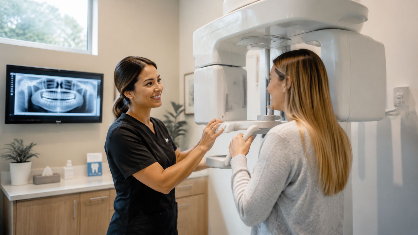
What the appointment usually feels like
If you need close-up images, a team member places a small sensor in your mouth for a few seconds at a time. You may need to bite gently to hold it in position. It's brief, and your dental assistant will guide you through each step.
If you need a panoramic image, you'll stand while the machine moves around your head. Nothing touches you beyond the positioning supports, and the scan is over quickly.
Patients often expect the process to feel more complicated than it really is. In most cases, it's one of the easiest parts of the visit.
What happens right after the image is taken
The biggest difference from older film systems is what happens next. Instead of waiting around, you and your dentist can often review the image almost immediately.
That makes the visit more interactive. Your dentist can point to a filling edge, an area between teeth, or a region near the root and explain what they're watching. If you're coming in for a first visit, you can learn more about what happens during a dental exam and how imaging fits into the full appointment.
Patients usually feel less anxious when they can see what the dentist sees and ask questions in real time.
Here's a short look at the experience many patients find helpful:
Questions patients often ask
Some patients worry they'll gag. Others worry about standing still too long or not knowing what they're supposed to do. A good dental team talks you through it as you go.
A few common concerns come up often:
- If you have dental anxiety: let the team know before the appointment begins. Small adjustments in pacing and communication can help.
- If you have jaw soreness or sensitivity: your assistant can work carefully and explain positioning before each image.
- If you're in pain: imaging is often one of the fastest ways to help your dentist find the cause and relieve uncertainty.
The visit usually feels less like a test and more like a guided conversation.
Schedule Your New Patient Exam with Dr. Schneider
If you want a dental office that combines clear communication with modern tools, digital imaging is one of the signs to look for. It supports safer, more efficient diagnosis, and it helps make treatment recommendations easier to understand.
For patients in Walnut Creek, CA, that can make a real difference during routine exams, urgent visits, cosmetic consultations, and restorative planning. Whether you're due for cleaning and exams, dealing with a painful tooth, considering tooth extraction, or looking for a long-term dental home, a modern imaging process can make your care feel more comfortable from the start.

Dr. Schneider's office welcomes new patients and families from Walnut Creek and the East Bay. If you've been searching for a dentist near me, dentist in Walnut Creek, CA, emergency dentist, or a practice that offers thoughtful preventive, cosmetic, and restorative dentistry, this is a good time to take the next step.
You can contact the office to schedule a new patient exam, ask about current concerns, or discuss treatment options such as dental implants, crowns, root canals, and cosmetic dentistry. The practice is located at 1855 San Miguel Dr., Suite 31, Walnut Creek, CA.
If you're ready for modern, patient-centered dental care, schedule a visit with William M. Schneider, DDS. You can reach the office to book a new patient exam, request a consultation, or get help for a pressing dental concern.